The Epidemiology, Pathogenesis, and Treatment of Type 1 Diabetes Mellitus
By
2015, Vol. 7 No. 11 | pg. 1/2 | »
IN THIS ARTICLE
KEYWORDS
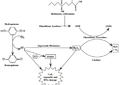
AbstractType 1 diabetes mellitus (T1DM), also known as insulin-dependent diabetes, is a chronic disease caused by autoimmune (type 1a) or spontaneous (type 1b) destruction of pancreatic beta cells, resulting in insulin deficiency. It is generally diagnosed in children before 20 years of age and is oftentimes fatal. This review will discuss the epidemiology of T1DM, including its incidence and prevalence, related temporal trends and risk factors for development. Furthermore, pathogenesis and immune system involvement of the disease will be evaluated, with a particular focus on cells of the adaptive and innate immune systems. Finally, an overview of past, present and future treatments for T1DM will be discussed. Type 1 diabetes mellitus (T1DM), also known as insulin-dependent diabetes, is a chronic disease caused by autoimmune (type 1a) or spontaneous (type 1b) destruction of pancreatic beta cells, resulting in insulin deficiency (Haller, Atkinson & Schatz, 2005) (Kim & Lee, 2009). A lack of insulin causes hyperglycaemia (high blood glucose levels), resulting in recurrent urination (polyuria); augmented thirst (polydipsia) and hunger (polyphagia); and weight loss (Haller, Atkinson & Schatz, 2005) (Gan, Albanese-O’Neill & Haller, 2012). Accounting for 10% of total diabetes cases, T1DM affects millions of individuals worldwide (Kim & Lee, 2009) (Achenbach, Bonifacio & Ziegler, 2005). It is generally diagnosed in children before 20 years of age (Gan, Albanese-O’Neill & Haller, 2012) (Maahs, West, Lawrence et al., 2010). Furthermore, the Juvenile Diabetes Research Foundation International (JDRF Int.) predicts an increase in the incidence of the disease among youth in upcoming years (Kim & Lee, 2009). Unfortunately, T1DM is fatal without treatment (Gan, Albanese-O’Neill & Haller, 2012).EpidemiologyIncidence and Prevalence of T1DMMultiple registry studies, including the World Health Organization Multinational Project for Childhood Diabetes (DIAMOND Project) (The DIAMOND Project Group, 2006) and SEARCH for Diabetes in Youth (SEARCH) (Liese, D’Agostino, Hamman et al., 2006) (Dabelea, Mayer-Davis & Imperatore, 2010), have been conducted to investigate the incidence of T1DM worldwide (Maahs, West, Lawrence et al., 2010). Firstly, the DIAMOND Project, initiated in 1990, aimed to study the development of T1DM in children. From 1990-1994, the incidence of T1DM in children (ages 14 and younger) was reported to be 0.036% (36 per 100,000 per year) in Finland and Sardinia (highest incidence of T1DMM in the world) (Maahs, West, Lawrence et al., 2010) (The DIAMOND Project Group, 2006). Furthermore, it was observed that the prevalence of the disease increased as age increased in most groups. Accordingly, the highest incidence of T1DM (~0.060% in 2005) was seen in 10-14 year-olds (Maahs, West, Lawrence et al., 2010). The DIAMOND Project reported a variation in the occurrence of the disease by country (The DIAMOND Project Group, 2006). The lowest incidence of T1DM (<1/100,000 cases per year) was observed in China and South America. Conversely, the highest incidence of the disease (>20/100,000 cases per year) was reported in Finland, Canada, and New Zealand. The investigators attributed the variation between ethnic groups to genetic and environmental differences (Maahs, West, Lawrence et al., 2010) (The DIAMOND Project Group, 2006). Secondly, the SEARCH for Diabetes in Youth Study investigated the incidence of T1DM in Americans 20 years of age and younger (Liese, D’Agostino, Hamman et al., 2006) (Dabelea, Mayer-Davis & Imperatore, 2010). SEARCH evaluated the onset of diabetes by age, gender, and ethnicity (Liese, D’Agostino, Hamman et al., 2006) (Dabelea, Mayer-Davis & Imperatore, 2010). The SEARCH Study suggested that incidence rates for T1DM reached a peak in age ranges 5-9 years and 10-14 years. Incidence of T1DM was reported to be equal in females and males (Liese, D’Agostino, Hamman et al., 2006). Furthermore, prevalence of the disease was observed to be highest in Caucasian youth (Gan, Albanese-O’Neill & Haller, 2012) (Maahs, West, Lawrence et al., 2010). Temporal TrendsAn updated report from the DIAMOND Project investigated the prevalence of T1DM in children (14 years of age and younger) from 57 countries. From 1990-1999, the average annual increase in the occurrence of the disease was 2.8%. The authors attributed the increase to gene-environment interactions (The DIAMOND Project Group, 2006). Risk Factors for Development of T1DMGender, ethnicity, genetics and environmental influences contribute to the development of T1DM (Gan, Albanese-O’Neill & Haller, 2012) (Maahs, West, Lawrence et al., 2010) (Ikegami, Noso, Babaya et al., 2011). Firstly, T1DM is prevalent in youth 20 years of age and younger. The incidence rates of the disease increase from birth and peak at age 14. The prevalence of T1DM decreases after puberty and stabilizes in adulthood. The number of new cases of diabetes is markedly lower in adults than in children (Gan, Albanese-O’Neill & Haller, 2012) (Maahs, West, Lawrence et al., 2010). Secondly, it has been found that female and male children are equally affected with T1DM. However, certain studies have shown that males are disproportionally affected in regions with a high prevalence of T1DM while females are disproportionally affected in regions with a low prevalence of the disease (Maahs, West, Lawrence et al., 2010). Thirdly, in a follow-up experiment, the SEARCH Study reviewed the incidence of T1DM in five ethnic groups (non-Hispanic white, African American, Hispanic, Asian and Pacific Islander, and Navajo populations). The authors indicated that the prevalence of T1DM is highest in non-Hispanic white populations and lowest in Navajo groups. Finally, genetics and environmental factors (discussed below) influence the onset of T1DM (Maahs, West, Lawrence et al., 2010). Genetic Susceptibility FactorsGenetics contribute to the onset of T1DM. In 1998, the EURODIAB Ace Study Group found that T1DM is common in families with a history of the disease (Lévy-Marchal, Patterson & Green, 2001). Moreover, Redondo et al. and Hyttinen et al. demonstrated a high (40-60%) concordance of T1DM in monozygotic twins (Redondo, Jeffrey, Fain et al., 2008) (Hyttinen, Kaprio, Kinnunen et al., 2003) Understanding the genetic factors that underlie T1DM will clarify the causes and progression of the disease. Furthermore, identifying genes that confer susceptibility for T1DM will aid in the establishment of prevention and treatment methods for diabetes patients (Concannon, Rich & Nepom, 2009). While some diseases are due to single gene mutations, many autoimmune diseases result from an unlucky combination of many gene variants. Each gene variant on its own is a ‘good’ (useful) gene variant (an evolutionarily selected polymorphism). However, in certain instances, a particular combination of gene variants may not work well together in terms of the immune system’s ability to establish or maintain tolerance of self antigens. The candidate gene approach and genome-wide association studies (GWAS) (Ikegami, Noso, Babaya et al., 2011) have been used to uncover over 40 susceptibility loci in T1DM, including insulin-coding genes (E.g. INS); human leukocyte antigen (HLA); interleukin (IL)-2 receptor alpha (CD25), cytotoxic T lymphocyte antigen (CTLA)-4; and the tyrosine phosphatase PTPN22 (Ghazarian, Diana, Simoni et al., 2013). The INS gene and the HLA loci, in particular, show strong associations with T1DM. Insulin Gene-Related Pathway, INSInsulin, coded by the INS gene, is a key autoantigen targeted by autoimmune responses in T1DM (Concannon, Rich & Nepom, 2009) (Pugliese, 2005). Transcription of the hormone is controlled by binding of the transcription factor Purl to the INS promoter region element called VNTR (variable number of tandem repeats) (Pugliese, 2005). Studies have found that the VNTR is a main susceptibility determinant of T1DM. For instance, homozygosity for the short Class VNTR I alleles is found in 75-85% of T1DM patients compared to 50-60% of the general population (Pugliese, 2005). This suggests a link between Class VNTR I alleles and T1DM. Moreover, homozygosity for the longer Class III VNTR alleles is rarely observed in T1DM patients, indicating a dominant protective effect (Pugliese, 2005). Major T1DM Susceptibility Gene, HLAThe HLA gene, also called IDDM1 (insulin-dependent diabetes mellitus locus), confers a strong risk for T1DM in most ethnic groups. The diabetes associated HLA Class II DR and DQ alleles are found in approximately 40-50% of T1DM cases (Ikegami, Noso, Babaya et al., 2011) (Ghazarian, Diana, Simoni et al., 2013). Differences in alleles and haplotypes of the HLA genes exist in Western and Asian countries. For instance, the DR3 and DR4 haplotypes confer T1DM susceptibility in Caucasian populations (Ghazarian, Diana, Simoni et al., 2013). In Lebanese populations, however, the DR4 haplotype is neutral in T1DM inception (Ghazarian, Diana, Simoni et al., 2013). Thus, the distribution of T1DM susceptibility alleles and haplotypes partially explains differences in worldwide incidences of the disease (Ghazarian, Diana, Simoni et al., 2013). The presence of HLA susceptibility alleles does not trigger the development of T1DM with absolute certainty. Hence, it is likely that external factors contribute to the onset of the disease as well. For instance, the frequency of susceptibility HLA-DQ alleles is similar in children from Finland and Karelia, a region of Russia. However, the incidence of T1DM is six times higher in Finland than Karelia, suggesting a non-genetic contributor (Ikegami, Noso, Babaya et al., 2011). Furthermore, it has been found that the incidence of T1DM differs in Western and Eastern Germany, despite similar genetic backgrounds in both areas (Ikegami, Noso, Babaya et al., 2011). Therefore, it is possible that a combination of genetics and environmental factors induce T1DM. Environmental Factors and Infections Contributing to Type 1 DiabetesEnvironmental factors contribute to the onset of T1DM. Epidemiological studies have found associations between T1DM and socio-economic status (SES), dietary and nutritional habits, and pathogen exposure. Socioeconomic FactorsIn a 2001 study, Patterson et al. showed that developed countries (high SES) have a higher incidence of T1DM than poorer nations (low SES) (Patterson, Dahlquist, Soltesz et al., 2001). Westernized countries place a strong emphasis on cleanliness and invest in education and medical care (e.g. vaccinations and antibiotics), resulting in higher levels of hygiene than developing countries (Ghazarian, Diana, Simoni et al., 2013). High levels of hygiene limit the survival, proliferation, and spread of pathogens. According to the “Hygiene Hypothesis,” a cleaner lifestyle suppresses the natural immune system (Ghazarian, Diana, Simoni et al., 2013) (Akerblom & Knip, 1998). An untrained immune system may respond inappropriately to self, increasing the frequency of autoimmune diseases, including T1DM (Ghazarian, Diana, Simoni et al., 2013) (Akerblom & Knip, 1998). Dietary Habits/Nutritional FactorsDietary habits can influence the development of T1DM (Ikegami, Noso, Babaya et al., 2011) (Akerblom & Knip, 1998). Firstly, the early introduction of cow’s milk into the diet of an infant may trigger the disease. A 1999 study by Elliott et al. compared milk consumption in 14 countries. It was found that the incidence of T1DM increased with increasing consumption of the milk protein β-casein (A1 and B variants) (Elliot, Harris, Hill et al., 1999). Accordingly, Nordic countries with higher milk consumption rates had high levels of T1DM while Iceland with lower milk consumption rates had low levels of T1DM (Ikegami, Noso, Babaya et al., 2011). Secondly, dietary gluten has been associated with the development of T1DM. A study of 1,600 children with T1DM parents found that the consumption of gluten before 3 months of age increases islet autoantibody risk (Grieco, Vendrame, Spagnuolo et al., 2011). Finally, nutritional factors, particularly levels of sun exposure and/or Vitamin D intake, contribute to T1DM inception. Vitamin D may have a protective effect against T1DM due to its immunosuppressive properties (Ghazarian, Diana, Simoni et al., 2013). Hence, countries with greater levels of sunlight and enhanced Vitamin D synthesis show a lower incidence of the disease. Furthermore, it has been reported that Vitamin D supplements during pregnancy reduce the risk of T1DM (Maahs, West, Lawrence et al., 2010) (Ghazarian, Diana, Simoni et al., 2013). It should be noted, however, that the above studies on diet are controversial, with other research showing no effect. Pathogenic Factors (Viruses and Enteroviruses)Viral infections may contribute to the development of T1DM via multiple mechanisms (Grieco, Vendrame, Spagnuolo et al., 2011). By mimicking sequence homology of a self-peptide, a pathogen-derived peptide may trigger an immune response against self-tissue in the host organism (known as molecular mimicry) (Grieco, Vendrame, Spagnuolo et al., 2011). Furthermore, viral infections may provoke inflammation and destruction of host cells, causing release of autoantigens and activation of autoreactive T cells (called bystander activation of T cells) (Grieco, Vendrame, Spagnuolo et al., 2011). Significant inflammation may induce stress in the endoplasmic reticulum (ER), resulting in protein denaturation and presentation of new autoantigens (known as antigen spreading) (Grieco, Vendrame, Spagnuolo et al., 2011). Secondly, human enteroviruses, including polioviruses; echoviruses; and rhinoviruses, are associated with T1DM. Enteroviruses are generally transmitted through consumption of contaminated food and drink. The rarity of enteroviruses in most developed countries is correlated with an increase in the incidence of T1DM, supporting the “Hygiene Hypothesis” (Luo, Herold & Miller, 2010). Pathogenesis of T1DM and Immune System InvolvementThe development of T1DM comprises a complex set of events from the activation of antigen-presenting cells (APCs) that present β-cell antigens to islet destruction and insulin deficiency (Kim & Lee, 2009). Pathogenesis of T1DM can be categorized into two stages. During the first stage (called insulitis), pancreatic islets are penetrated by leukocytes (Kim & Lee, 2009). After a benign prelude, insulin-producing β-cells are destroyed (Kim & Lee, 2009). The immune system comprises innate and adaptive immunity (Kim & Lee, 2009) (Wen, Ley, Volchkov et al., 2008). The adaptive and innate immune system uses Mϕs, DCs, NK, NK T, γδ T, αβT, and β cells to recognize dangerous molecules and induce an immediate immune response (Wen, Ley, Volchkov et al., 2008) (Janeway & Medzhitov, 2002). Moreover, cells of the innate immune system may activate antigen-specific cells of the adaptive immune system (Kim & Lee, 2009) (Wen, Ley, Volchkov et al., 2008). Recent studies have found that the innate immune system is associated with the development of autoimmune responses in inflammatory environments (Kim & Lee, 2009) (Zeigler & Nepom, 2010). In healthy tissues, APCs, including Mϕs and dendritic cells (DCs), are activated in response to infected host cells (Kim & Lee, 2009). By presenting self-antigens to autoreactive T cells, APCs can remove damaged cells. In inflamed tissues, however, signals that encourage APC maturation are prematurely generated. Hence, self-antigen-specific T and B cells are activated, causing an autoimmune response (Kim & Lee, 2009). Adaptive and innate immune cells, including Mϕs, DCs, NK, NK T γδ T, αβT, and β cells, are crucial to the pathogenesis of T1DM. MϕsMϕs are vital for host defence. Using pattern recognition receptors (PRRs), Mϕs can recognize pathogen-associated molecular patterns (PAMPs) and can activate immune regulators (Kim & Lee, 2009) (Akira, Uematsu & Takeuchi, 2006). However, they can cause extensive local damage when uncontrolled. In T1DM mice, Mϕs invade pancreatic islets before NK and autoreactive T- and B-cells (Kim & Lee, 2009). Furthermore, Mϕs from NOD mice (T1DM animal model) produce higher levels of proinflammatory cytokines (E.g. IL-12, TNFα, and IL-1α), promoting the differentiation of diabetogenic cells (Kim & Lee, 2009). Finally, Mϕs have been implicated in the direct destruction of B-cells (Kim & Lee, 2009). Hence, Mϕs contribute to the pathogenesis of T1DM. Dendritic Cells (DCs)Dendritic cells (DCs) are crucial stimulators of the adaptive immune system (Grieco, Vendrame, Spagnuolo et al., 2011). DCs accumulate in pancreatic islets of T1DM patients and NOD mice. In NOD mice, a subset of DCs receives maturation signals (Grieco, Vendrame, Spagnuolo et al., 2011). Mature DCs are responsible for capturing ß-cell antigens from islet cells and priming diabetogenic T cells (Grieco, Vendrame, Spagnuolo et al., 2011). Hence, DCs contribute to the onset of the disease. NK CellsNK cells are responsible for attacking host cells that are infected by foreign microorganisms (Kim & Lee, 2009) (Dotta, Fondelli & Falorni, 2008). Furthermore, they are potent moderators of APCs, including Mϕs and DCs (Grieco, Vendrame, Spagnuolo et al., 2011).It has been hypothesized that NK cells may promote or protect against T1DM depending on anatomical location and chemical environment. However, the data are inconclusive (Dotta, Fondelli & Falorni, 2008). Research has shown that NK cells can mediate the breakdown of pancreatic islet cells, contributing to the onset of the disease (MacKay, Jacobson & Rabinovitch, 1986). Furthermore, reduction of NK cells in NOD mice suppresses the progression of induced T1DM, suggesting that NK cells influence T1DM development (Kim & Lee, 2009). Conversely, studies have indicated that NK cells protect against T1DM. Impaired function of NK cells in T1DM patients suggests a protective function of NK cells (Grieco, Vendrame, Spagnuolo et al., 2011). NK T CellsNK T cells, a subset of T cells, are regulatory cells that have the ability to secrete IL-4 (Kim & Lee, 2009). NK T cells can recognize glycolipid antigens, includingα-galactosylceramide. NK T cells may protect against T1DM (Grieco, Vendrame, Spagnuolo et al., 2011). In NOD mice, activation of NK T cells by treatment withα-galactosylceramide provides protection against T1DM (Grieco, Vendrame, Spagnuolo et al., 2011). γδ T Cellsγδ T cells are crucial for linking the innate and the adaptive immune systems (Kim & Lee, 2009). They are not MHC restricted and can recognize a broader range of antigens compared to αβ T cells. γδ T cells can detect molecules produced by microorganisms. Furthermore, they can recognize self-antigens expressed in epithelial cells that have experienced tissue stress and damage (Kim & Lee, 2009). γδ T cells are involved in the development of autoimmune diseases, including T1DM. Studies have shown that γδ T cells are found in areas of inflammation due to autoimmune diseases (Kim & Lee, 2009). Furthermore, research has found that numbers of γδ T cells are reduced in NOD mice and T1DM patients (Kim & Lee, 2009). αβ T CellsThe role of T cells in T1DM is well established. Both CD4+ and CD8+ cells play a pathogenic role in mediating the development of T1DM. Under normal circumstances, T cells (i.e. white blood cells) protect the body from a variety of illnesses. However, in T1DM, T cells receive a signal to destroy β cells in the insulin-producing areas of the pancreas. It is not entirely certain why or how the signal is issued. Understanding the development of rogue T cells will better allow scientists and clinicians to treat T1DM (Wagner, 2011). β CellsInsulin-producing β cells in the pancreatic islets of Langerhans are important in the development of T1DM, particularly in disease onset and progression. β cells serve as antigen-presenting cells and autoantibody secretors in the lead up to T cell-mediated autoimmune destruction of insulin-producing β cells in T1DM. Consequently, there is significant interest in the use of β cell depletion therapies as a treatment for T1DM (Ekici, 2010). Experimental ModelsAnimal Models (Mice and Rats)Animal models have improved our understanding of the immunological mechanisms that underlie T1DM. Moreover, animal models are crucial for developing therapeutic interventions for diabetes patients. Two commonly used animal models for T1DM include the NOD mouse and the BioBreeding (BB) rat (Kim & Lee, 2009). The NOD mouse and the BB rat develop T1DM by autoreactive ab T cell-mediated destruction of pancreatic ß-cells (Kim & Lee, 2009). The progression of T1DM in NOD mice is similar to the progression of the disease in humans (Anderson & Bluestone, 2005). However, the incidence rate of T1DM in female and male NOD mice differs from the incidence rate of T1DM in female and male patients (Kim & Lee, 2009). For example, the incidence of T1DM in female NOD mice is 60-80% while the incidence of the disease in male NOD mice is 20-30% (Kim & Lee, 2009). In contrast, the incidence of T1DM is equal in women and men (Gan, Albanese-O’Neill & Haller, 2012). The pattern of T1DM development in BB rats is similar to the pattern of the disease in humans. However, BB rats experience T cell lymphopenia during T1DM, unlike human subjects (Kim & Lee, 2009). Clinical Trials and Clinical ResearchClinical trials are a crucial research tool used to advance medical knowledge about T1DM (Gan, Albanese-O’Neill & Haller, 2012). They can be used to investigate T1DM under different conditions. By studying the effects of different environmental factors on the development of T1DM, researchers can attain a stronger understanding of the underlying mechanisms of the disease. They may then investigate therapies that diminish the effects of T1DM (Gan, Albanese-O’Neill & Haller, 2012). In 2002, a clinical trial tested the glycaemic control of patients who use continuous subcutaneous insulin infusion and patients who use intensive insulin injections (Ikegami, Noso, Babaya et al., 2011). It was found that glycaemic control was higher in patients who used continuous subcutaneous insulin infusion compared to patients who used intensive insulin injections. Furthermore, it was discovered that less insulin was needed to achieve a strict control level in patients who used continuous subcutaneous insulin infusion (Ikegami, Noso, Babaya et al., 2011). It is clear that clinical trials are frequently used to investigate multiple aspects of T1DM.Continued on Next Page » 
From the Inquiries Journal Blog   Related ReadingMonthly Newsletter SignupThe newsletter highlights recent selections from the journal and useful tips from our blog. Suggested Reading from Inquiries Journal
Inquiries Journal provides undergraduate and graduate students around the world a platform for the wide dissemination of academic work over a range of core disciplines. Representing the work of students from hundreds of institutions around the globe, Inquiries Journal's large database of academic articles is completely free. Learn more | Blog | Submit Follow IJ
Latest in Health Science |