|
Featured Article: Assessing the Impact of Adverse Childhood Experiences on Brain Development
Children exposed to conditions involving abuse or neglect are faced with atypical caregiving at periods in development when the brain is markedly more sensitive to input from the environment. Childhood maltreatment is a broad dimension that encompasses physical, emotional, and sexual abuse, as well as any kind of physical or emotional neglect. Conditions involving abuse expose children to increased levels of traumatic input, whereas conditions of neglect are missing critical developmental experiences.
The limbic system supports a wide variety of functions and so continued efforts to better understand how maltreatment impacts the developmental outcomes of the amygdala and hippocampus have been particularly important. Early exposure to abuse and neglect have been linked to greater rates of pediatric posttraumatic stress disorder (PTSD).19 In a previous study, the use of anatomical imaging showed that more pronounced differences in the brain structures of youth with maltreatment-related PTSD were associated with an earlier age of onset, length of abuse duration, and increased PTSD symptoms relative to non-maltreated youth.1
Maltreated youth without PTSD exhibited larger amygdala and hippocampal volumes relative to youth with PTSD and non-maltreated controls. 2 This observation is consistent with previous findings in which maltreated PTSD youth had no significant differences in amygdala and hippocampal volumes compared to non-maltreated controls. 1 Substantial psychopathology for internalizing disorders and externalizing problems was observed in pediatric patients with maltreatment-related PTSD. 2
Smaller posterior cerebral and cerebellar grey matter volumes were observed in maltreated PTSD youth relative to maltreated youth without PTSD and non-maltreated controls.2 The neurobiological substrate by which volumetric decreases in posterior cortex and cerebellar grey matter may be influenced by an association between trauma mechanisms and an oversensitivity to PTSD symptoms that drives comorbidity.19
These posterior cortical regions support face recognition, language, and object recognition, and diffuse projections to frontal and parietal cortices and subcortical structures are involved in visuospatial imagery, retrieval of episodic memories, and self-processing operations.2 It has been discussed that grey matter deficits in these regions have the potential to mitigate the formation of lucid memories about the traumatic experiences, driving the impaired processing of traumatic cues and the inability to abrogate PTSD symptoms.2
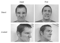
In one study that used the fMRI technique, increased activation of the anterior insula (AI) and amygdala was observed in response to angry faces but not sad faces in children who were previously exposed to family violence.6 Furthermore, the family violence group showed heightened activity in the right amygdala and bilaterally in the AI when angry faces but not sad faces were presented with neutral faces.6 The greater activation of the right amygdala but not the left amygdala corroborates the theory that emotional perception and expression are lateralized to the right hemisphere.
Children exposed to maltreatment often exhibit deficits in executive functioning. Several studies have employed electrophysiological approaches to assess any functional alterations during cognitive tasks resulting from maltreatment. Interestingly, tasks that rely on medial temporal areas and those that rely on frontal cortex have a more pronounced decrement in performance.3
In one study focusing on the impact of socioeconomic status (SES) on brain function, evidence supported that SES is significantly associated with neurocognitive development.3 The most extreme SES disparities were observed in language and memory ability, followed by working memory ability, and then trended more weakly towards varying levels of ability for cognitive control.3 Despite being a sociological construct, SES encompasses an extremely broad domain of physical and psychosocial factors that will ultimately shape brain development.
One study expanded upon the current body of research by examining the impact of developmental timing of maltreatment on higher-order functioning and showed increased deficits in performance on both inhibitory control and working memory tasks in maltreated youth compared to controls.21 Furthermore, it provided evidence that children who were either maltreated as infants or have a history of maltreatment performed more poorly than children without a chronic history of maltreatment.21 This adds credence to the idea that exposure to maltreating environments during infancy induces structural and functional deviations. The protracted stress of chronic maltreatment during infancy further potentiates these deficits.21
Well-timed interventions are particularly important for young children who are most vulnerable to early physiological disruptions that alter normal structural and functional brain development. There is substantial evidence supporting Trauma-Focused Cognitive-Behavioral Therapy as an effective treatment to reduce symptoms of PTSD and depression, as well as behavioral difficulties in children who have experienced sexual abuse, domestic violence, or other childhood traumas.11 Evidence of decreased behavioral problems in foster children in response to treatment with parent-child interaction therapy (PCIT) has been documented across several studies.12,13
The primary insight gained from experimental evidence is that early exposure to adversity disrupts normative brain development. Findings across studies have indicated that deficits in developmental structure and function are further instantiated by cumulative exposures to adverse rearing experiences.21 Time-dependent and region-specific sensitive periods drive the protracted course of brain development.10 There is strong evidence that well-timed therapeutic intervention can abrogate changes in developmental trajectories.11,12,13
Promising new initiatives to change the current face of primary health care make this an exciting time to be involved in research that is currently at the interface between science and medicine. As the present body of knowledge on brain development expands, so will the capacity to effectively contribute towards the resolve of this public health concern.
- De Bellis, M. D., M. S. Keshavan, H. Shifflett, S. Iyengar, S. R. Beers, J. Hall, and G. Moritz. “Brain Structures in Pediatric Maltreatment-Related Posttraumatic Stress Disorder: A Sociodemographically Matched Study.” Biological Psychiatry 52.11 (2002): 1066-078. Elsevier. Web. 10 Mar. 2016.
- De Bellis, Michael D., Stephen R. Hooper, Steven D. Chen, James M. Provenzale, Brian D. Boyd, Christopher E. Glessner, James R. MacFall, Martha E. Payne, Robert Rybczynski, and Donald P. Woolley. “Posterior Structural Brain Volumes Differ in Maltreated Youth with and without Chronic Posttraumatic Stress Disorder.” Development and Psychopathology 27 (2015): 1555-576. Web. 13 Mar. 2016.
- Farah, Martha J., David M. Shera, Jessica H. Savage, Laura Betancourt, Joan M. Giannetta, Nancy L. Brodsky, Elsa K. Malmud, and Hallam Hurt. “Childhood Poverty: Specific Associations with Neurocognitive Development.” Brain Research 1110.1 (2006): 166-74. Elsevier. Web. 1 Mar. 2016.
- Hanson, Jamie L., Brendon M. Nacewicz, Matthew J. Sutterer, Amelia A. Cayo, Stacey M. Schaefer, Karen D. Rudolph, Elizabeth A. Shirtcliff, Seth D. Pollak, and Richard J. Davidson. “Behavioral Problems after Early Life Stress: Contributions of the Hippocampus and Amygdala.” Biological Psychiatry 77 (2014): 314-23. Web. 13 Mar. 2016.
- Hodel, Amanda S., Ruskin H. Hunt, Raquel A. Cowell, Sara E. Van Den Heuvel, Megan R. Gunnar, and Kathleen M. Thomas. “Duration of Early Adversity and Structural Brain Development in Post-Institutionalized Adolescents.” NeuroImage 105 (2015): 112-19. Elsevier. Web. 12 Mar. 2016.
- McCrory, Eamon J., Stephane A. De Brito, Catherine L. Sebastian, Andrea Mechelli, Geoffrey Bird, Phillip A. Kelly, and Essi Viding. “Heightened Neural Reactivity to Threat in Child Victims of Family Violence.” Current Biology 21.23 (2011): 947-48. Elsevier. Web. 13 Mar. 2016.
- Tottenham, N., T. A. Hare, A. Millner, J.D. Zevin, and B. J. Casey. “Elevated Amygdala Response to Faces following Early Deprivation.” Developmental Science 14.2 (2011): 190-204. Web. 13 Mar. 2016.
- Tottenham, Nim, and Margaret A. Sheridan. “A Review of Adversity, The Amygdala and the Hippocampus: A Consideration of Developmental Timing.” Frontiers in Human Neuroscience 3 (2010): 1-18. PMC. Web. 10 Mar. 2016.
- White, Michael G., Ryan Bogdan, Patrick M. Fisher, Karen Munoz, Douglas E. Williamson, and Ahmad R. Hariri. “FKBP5 and Emotional Neglect Interact to Predict Individual Differences in Amygdala Reactivity.” Genes, Brain, and Behavior 11.7 (2012): 869-78. PMC. Web. 5 Mar. 2016.
- Wieranga, Lara M., Martjin P. Van Den Heuvel, Sarai Van Dijk, Yvonne Rijks, Marcel A. De Reus, and Sarah Durston. “The Development of Brain Network Architecture.” Human Brain Mapping 37 (2016): 717-29. Print.
- “Trauma-Focused Cognitive Behavioral Therapy for Children Affected by Sexual Abuse or Trauma.” Child Welfare. Child Welfare Information Gateway, Aug. 2012. Web. 25 Mar. 2016.
- Timmer, Susan, Anthony Urquiza, and Nancy Zebell. “Challenging Foster Caregiver-Maltreated Child Relationships: The Effectiveness of Parent-Child Interaction Therapy.” Children and Youth Services Review 28 (2006): 1-19. Elsevier. Web. 2 Apr. 2016.
- McNeil, Cheryl, Amy Herschell, Robin Gurwitch, and Laurie Clemens-Mowrer. “Training Foster Parents in Parent-Child Interaction Therapy.” Education and Treatment of Children 28.2 (2005): 182-96. JSTOR. Web. 2 Apr. 2016.
- Bruce, Jacqueline, Philip Fisher, Alice Graham, William Moore, III, Shannon Peake, and Anne Mannering. “Patterns of Brain Activation in Foster Children and Nonmaltreated Children during an Inhibitory Control Task.” Development and Psychopathology 25 (2013): 931-41. Cambridge University Press. Web. 2 Apr. 2016.
- Aron, A. R., P. C. Fletcher, E. T. Bullmore, B. J. Sahakian, and T. W. Robbins. “Stop-Signal Inhibition Disrupted by Damage to Right Inferior Frontal Gyrus in Humans.” Nature Neuroscience 6 (2003): 115-16. NCBI. Web. 2 Apr. 2016.
- Banich, Marie T., and Rebecca J. Compton. Cognitive Neuroscience. 3rd ed. Belmont, CA: Wadsworth, Cengage Learning, 2011. Print.
- Cook, S. C., and C. L. Wellman. “Chronic Stress Alters Dendritic Morphology in Rat Medial Prefrontal Cortex.” Journal of Neurobiology. 60.2 (2004): 236-48. NCBI. Web. 2 Apr. 2016.
- Tottenham, Nim, et al. “Prolonged Institutional Rearing is Associated with Atypically Larger Amygdala Volume and Difficulties in Emotion Regulation.” Developmental Science 13.1 (2010): 46. PMC. Web. 3 Apr. 2016.
- DeBellis, M. D., and A. Zisk. “The Biological Effects of Childhood Trauma.” Child and Adolescent Psychiatric Clinics of North America: Disaster and Trauma 23.2 (2014): 185-222. NIH. Web. 2 Apr. 2016.
- Hodges, Jill, and Barbara Tizard. “Social and Family Relationships of Ex-Institutional Adolescents.” Journal of Child Psychology and Psychiatry 30.1 (1989): 77-97. Wiley. Web. 3 Apr. 2016.
- Cowell, Raquel, Dante Cicchetti, Fred Rogosch, and Sheree Toth. “Childhood Maltreatment and its Effect on Neurocognitive Functioning: Timing and Chronicity Matter.” Development and Psychopathology 27 (2015): 521-33. Cambridge University Press. Web. 2 Apr. 2016.
Endnotes
- De Bellis, M. D., M. S. Keshavan, H. Shifflett, S. Iyengar, S. R. Beers, J. Hall, and G. Moritz. “Brain Structures in Pediatric Maltreatment-Related Posttraumatic Stress Disorder: A Sociodemographically Matched Study.” Biological Psychiatry 52.11 (2002): 1066-078. Elsevier. Web. 10 Mar. 2016.
- De Bellis, Michael D., Stephen R. Hooper, Steven D. Chen, James M. Provenzale, Brian D. Boyd, Christopher E. Glessner, James R. MacFall, Martha E. Payne, Robert Rybczynski, and Donald P. Woolley. “Posterior Structural Brain Volumes Differ in Maltreated Youth with and without Chronic Posttraumatic Stress Disorder.” Development and Psychopathology 27 (2015): 1555-576. Web. 13 Mar. 2016.
- Farah, Martha J., David M. Shera, Jessica H. Savage, Laura Betancourt, Joan M. Giannetta, Nancy L. Brodsky, Elsa K. Malmud, and Hallam Hurt. “Childhood Poverty: Specific Associations with Neurocognitive Development.” Brain Research 1110.1 (2006): 166-74. Elsevier. Web. 1 Mar. 2016.
- Hanson, Jamie L., Brendon M. Nacewicz, Matthew J. Sutterer, Amelia A. Cayo, Stacey M. Schaefer, Karen D. Rudolph, Elizabeth A. Shirtcliff, Seth D. Pollak, and Richard J. Davidson. “Behavioral Problems after Early Life Stress: Contributions of the Hippocampus and Amygdala.” Biological Psychiatry 77 (2014): 314-23. Web. 13 Mar. 2016.
- Hodel, Amanda S., Ruskin H. Hunt, Raquel A. Cowell, Sara E. Van Den Heuvel, Megan R. Gunnar, and Kathleen M. Thomas. “Duration of Early Adversity and Structural Brain Development in Post-Institutionalized Adolescents.” NeuroImage 105 (2015): 112-19. Elsevier. Web. 12 Mar. 2016.
- McCrory, Eamon J., Stephane A. De Brito, Catherine L. Sebastian, Andrea Mechelli, Geoffrey Bird, Phillip A. Kelly, and Essi Viding. “Heightened Neural Reactivity to Threat in Child Victims of Family Violence.” Current Biology 21.23 (2011): 947-48. Elsevier. Web. 13 Mar. 2016.
- Tottenham, N., T. A. Hare, A. Millner, J.D. Zevin, and B. J. Casey. “Elevated Amygdala Response to Faces following Early Deprivation.” Developmental Science 14.2 (2011): 190-204. Web. 13 Mar. 2016.
- Tottenham, Nim, and Margaret A. Sheridan. “A Review of Adversity, The Amygdala and the Hippocampus: A Consideration of Developmental Timing.” Frontiers in Human Neuroscience 3 (2010): 1-18. PMC. Web. 10 Mar. 2016.
- White, Michael G., Ryan Bogdan, Patrick M. Fisher, Karen Munoz, Douglas E. Williamson, and Ahmad R. Hariri. “FKBP5 and Emotional Neglect Interact to Predict Individual Differences in Amygdala Reactivity.” Genes, Brain, and Behavior 11.7 (2012): 869-78. PMC. Web. 5 Mar. 2016.
- Wieranga, Lara M., Martjin P. Van Den Heuvel, Sarai Van Dijk, Yvonne Rijks, Marcel A. De Reus, and Sarah Durston. “The Development of Brain Network Architecture.” Human Brain Mapping 37 (2016): 717-29. Print.
- “Trauma-Focused Cognitive Behavioral Therapy for Children Affected by Sexual Abuse or Trauma.” Child Welfare. Child Welfare Information Gateway, Aug. 2012. Web. 25 Mar. 2016.
- Timmer, Susan, Anthony Urquiza, and Nancy Zebell. “Challenging Foster Caregiver-Maltreated Child Relationships: The Effectiveness of Parent-Child Interaction Therapy.” Children and Youth Services Review 28 (2006): 1-19. Elsevier. Web. 2 Apr. 2016.
- McNeil, Cheryl, Amy Herschell, Robin Gurwitch, and Laurie Clemens-Mowrer. “Training Foster Parents in Parent-Child Interaction Therapy.” Education and Treatment of Children 28.2 (2005): 182-96. JSTOR. Web. 2 Apr. 2016.
- Bruce, Jacqueline, Philip Fisher, Alice Graham, William Moore, III, Shannon Peake, and Anne Mannering. “Patterns of Brain Activation in Foster Children and Nonmaltreated Children during an Inhibitory Control Task.” Development and Psychopathology 25 (2013): 931-41. Cambridge University Press. Web. 2 Apr. 2016.
- Aron, A. R., P. C. Fletcher, E. T. Bullmore, B. J. Sahakian, and T. W. Robbins. “Stop-Signal Inhibition Disrupted by Damage to Right Inferior Frontal Gyrus in Humans.” Nature Neuroscience 6 (2003): 115-16. NCBI. Web. 2 Apr. 2016.
- Banich, Marie T., and Rebecca J. Compton. Cognitive Neuroscience. 3rd ed. Belmont, CA: Wadsworth, Cengage Learning, 2011. Print.
- Cook, S. C., and C. L. Wellman. “Chronic Stress Alters Dendritic Morphology in Rat Medial Prefrontal Cortex.” Journal of Neurobiology. 60.2 (2004): 236-48. NCBI. Web. 2 Apr. 2016.
- Tottenham, Nim, et al. “Prolonged Institutional Rearing is Associated with Atypically Larger Amygdala Volume and Difficulties in Emotion Regulation.” Developmental Science 13.1 (2010): 46. PMC. Web. 3 Apr. 2016.
- DeBellis, M. D., and A. Zisk. “The Biological Effects of Childhood Trauma.” Child and Adolescent Psychiatric Clinics of North America: Disaster and Trauma 23.2 (2014): 185-222. NIH. Web. 2 Apr. 2016.
- Hodges, Jill, and Barbara Tizard. “Social and Family Relationships of Ex-Institutional Adolescents.” Journal of Child Psychology and Psychiatry 30.1 (1989): 77-97. Wiley. Web. 3 Apr. 2016.
- Cowell, Raquel, Dante Cicchetti, Fred Rogosch, and Sheree Toth. “Childhood Maltreatment and its Effect on Neurocognitive Functioning: Timing and Chronicity Matter.” Development and Psychopathology 27 (2015): 521-33. Cambridge University Press. Web. 2 Apr. 2016.
Suggested Reading from Inquiries Journal
Post-traumatic stress disorder in children under six years old has been formally recognized since 2013 (Veteran’s Affairs, 2019), yet the body of research is still lacking for this age group. An important step towards helping these youngest sufferers of post-traumatic stress disorder is to determine whether symptomology assessments... MORE»
This paper presents and evaluates the varying roadblocks that make identifying and assessing emotional abuse to children so complex. This is the case for three primary reasons: the lack of a common definition of what constitutes... MORE»
Conversations about the gender expression of young children are often characterized by confusion, as parents, educators, and even child psychologists have a hard time determining where exactly children’s strong gendered beliefs and behaviors come from. Children as young as two have been observed exhibiting behaviors and expressing... MORE»
For today's children, technological devices such as iPads, smartphones, and e-readers are quickly replacing more traditional "toys" as sources of learning and entertainment. With their capacity to contain a multitude of activities within a single device, tech devices are the new norm and are being utilized from a young age. Many... MORE»
Latest in Neuroscience
2020, Vol. 12 No. 11
Isochronic tones are a hypothesized auditory brainwave entrainment technique in which a single tone is played at regular beat intervals. Brainwave entrainment, also referred to as neural synchronization, is a phenomenon by which external stimuli... Read Article »
2013, Vol. 5 No. 09
Antisocial personality disorder (ASPD), also known as dyssocial personality disorder, is a mental illness that is characterized by a reckless disregard for social norms, impulsive behaviour, an inability to experience guilt, and a low tolerance... Read Article »
2016, Vol. 8 No. 12
Huntington’s disease is a progressive neurodegenerative disorder that affects around five people in every 100,000. It is caused by an increase in a polyglutamine region of the Huntingtin protein, resulting in a toxic gain of function mutation... Read Article »
2015, Vol. 11 No. 1
Neurofeedback Therapy (NFT) is a type of biofeedback therapy specifically targeting the brain and nervous system. According to the Mayo Clinic, biofeedback is defined as a technique one can use to learn to control the body’s functions, done... Read Article »
2014, Vol. 6 No. 09
Autism is a complex neuro-developmental disorder causing deficits in social interaction and language development at an early age. The severity is based on the level of impaired social communication and restricted, repetitive behaviors. The average... Read Article »
2007, Vol. 2 No. 1
The peripheral nervous system is made up of the nerves and neurons that are outside of the central nervous system. These nerves and neurons are used to transport information between the brain and the rest of the body, and when damaged, can severely... Read Article »
2011, Vol. 3 No. 03
When investigating the effect of gaze direction on facial expressions of emotion, previous imaging research indicated that dynamic presentation of stimuli produced higher amygdala responses (Sato, Kochiyama, Uono, & Yoshikawa, 2010). A behavioral... Read Article »
|